Scientists Map Diaphragm’s Motor Nerve and Arteriolar Networks With Neurolucida
A 3D model of a mouse diaphragm appears on the monitor. Blood vessels branch out from entry points around the muscle’s periphery, engaging in a graceful choreography with the nerve fibers that radiate from its center.
Could these two networks work together to ensure healthy blood and oxygen flow to the muscle? Or do they exist independently of each other, house mates living side by side within the confines of the diaphragm? Dr. Diego Correa and Dr. Steven Segal set out to test the hypothesis that the motor innervation and blood supply of the diaphragm muscle are physically associated.
“We used Neurolucida to map entire arteriolar networks together with entire motor nerve networks of the diaphragm muscle in adult mice,” explained Dr. Segal in an email.
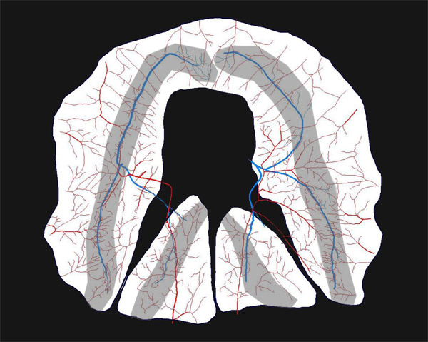
Image adapted from “Neurovascular proximity in the diaphragm muscle of adult mice,” published with permission from Dr. S. Segal
The scientists from Yale University and the University of Missouri filled arterioles with a casting compound and stained motor nerves so that each branch of the two networks could be visible using brightfield imaging.
“By mapping respective networks simultaneously within each muscle, Neurolucida enabled us to perform a proximity analysis to quantify the separation distance between motor nerves and arterioles,” Dr. Segal said. To measure this distance, they used Neurolucida to calculate the space between each micron along the nerve network and the nearest arteriole.
They found a consistent physical association between the two networks that, they posit, is not the result of random positioning; in fact, the proximity of the two networks might facilitate blood flow to the local regions within the muscle.
“Our findings suggest that neurovascular proximity within the diaphragm may facilitate vasoactive signals arising from motor unit recruitment to be ‘sensed’ by nearby arterioles,” the authors say in their paper “Neurovascular Proximity in the Diaphragm Muscle of Adult Mice” published in the journal Microcirculation.
Both strains of mice analyzed in the study showed an association between motor nerves and arterioles that, according to the authors, “are more closely associated with each other than can be explained by chance, providing a foundation for exploring how these relationships may influence blood flow control and whether they are altered with injury, disease or aging.”
Read more about the study at PubMed.gov
Correa D & Segal SS. Neurovascular proximity in the diaphragm muscle of adult mice. Microcirculation 19: 306–315, 2012. doi: 10.1111/j.1549-8719.2012.00163.x