Neuroscientists in Germany Outline Protocol for Best Accuracy in Neuron Reconstruction, including Use of Neurolucida
 No two neurons are exactly alike. Structure dictates function, so for scientists to fully understand the way different types of neurons work, they must first get to know their forms.
Scientists at the Institute for Neuroscience and Medicine at the Research Center Jülich in Jülich, Germany use Neurolucida to perform neuron reconstruction, the most effective method for studying neuron morphology.
In their paper “Improved biocytin labeling and neuronal 3D reconstruction,” published last year in Nature Protocols, the German team describes a distinct series of steps, which must be carried out before a truly accurate model of a neuron can be created. From brain dissection and slice preparation to fixation, staining, embedding, and 3D reconstruction, the authors clearly lay out the process.
In detailing their protocol, the team took into consideration common issues that occur with the embedding and labeling of neuronal tissue such as shrinkage, distortion, and fading. Biocytin labeling, they say, is superior to other methods because of the “extremely durable and strong staining” it achieves. According to them, the labeling method also allows for tissue to be re-examined “to test a new scientific hypothesis or to verify the findings in a different context.”
In one section of the protocol, entitled “Suggestions for 3D, light-microscopic reconstructions of neurons,” the authors describe how to perform 3D reconstructions of biocytin labeled neurons with Neurolucida. “This software allows manual reconstructions of neurons in all three dimensions and generates reconstruction data files in the Neurolucida format for a quantitative morphological analysis,” they explain.
Read “Improved biocytin labeling and neuronal 3D reconstruction” at nature.com.
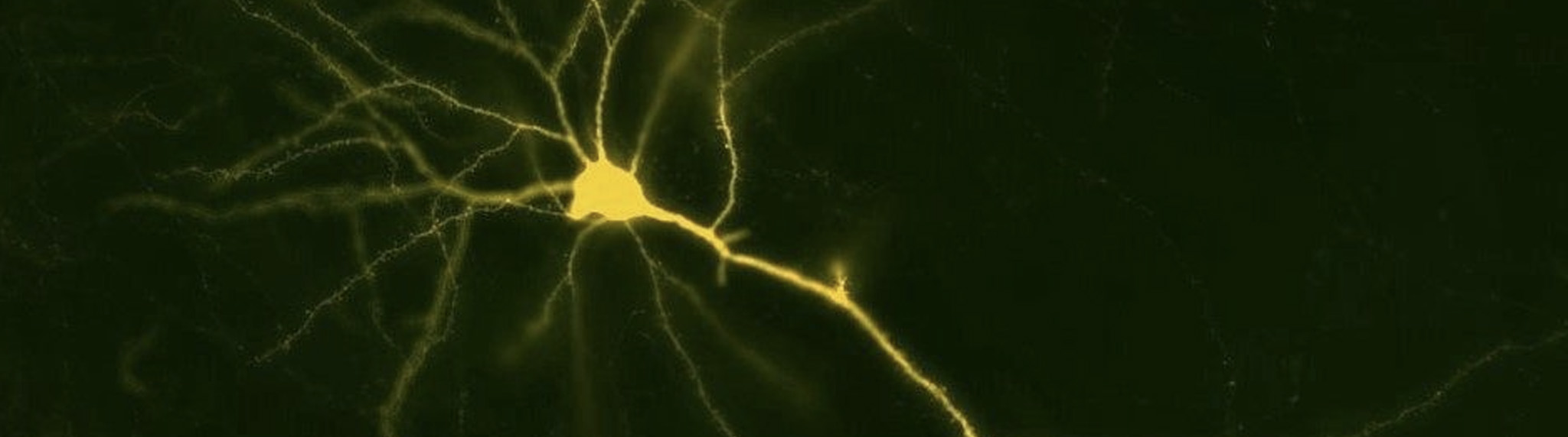
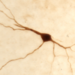
(Note that the image above is for illustration purposes only and was not actually used in the study described in this post.)
Marx, M., Günter, R. H., Hucko, W., Radnikow, G., & Feldmeyer, D. (2012). Improved biocytin labeling and neuronal 3D reconstruction. Nature protocols,7(2), 394-407.