UVM Scientists Use Neurolucida and Stereo Investigator to Study Neurons in the Avian Iris
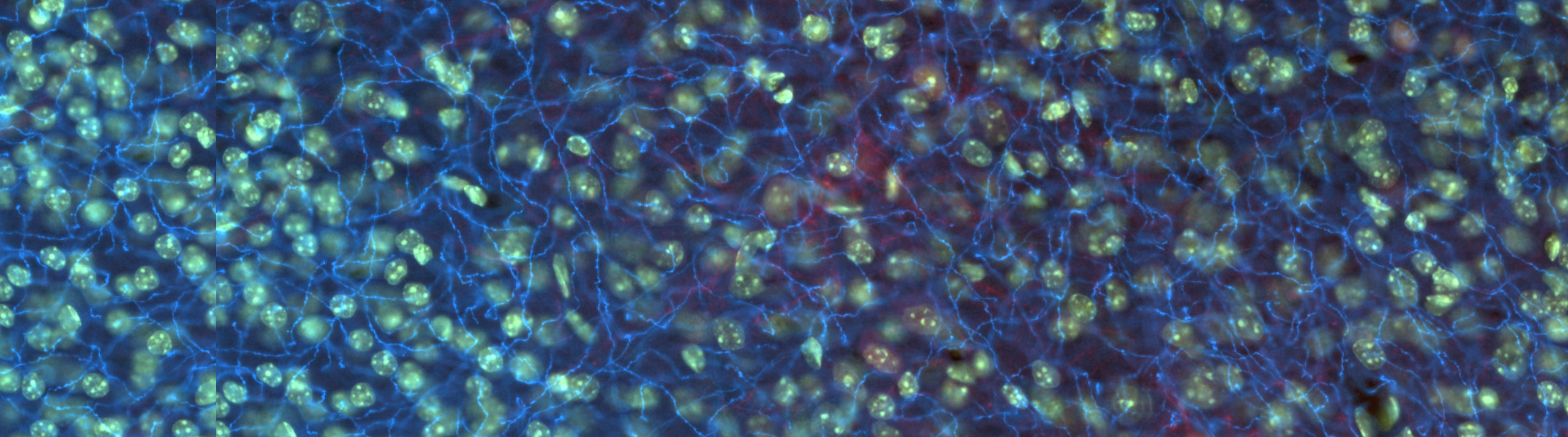
During a chicken embryo’s twenty-one days of incubation, its eyes develop in astonishing ways. Muscles form, neurons branch, innervation occurs. Researchers at Dr. Rae Nishi’s lab at the University of Vermont, including two MBF Bioscience staff scientists Julie Simpson, Ph.D. and Julie Keefe, M.S. are studying the development of a chicken embryo’s nervous system. Their specific focus is on the behavior of neurons in the ciliary ganglion – a mass of nerve cells in the eye’s ciliary muscle.
Published last month in Developmental Neurology, their paper “Differential effects of RET and TRKB on axonal branching and survival of parasympathetic neurons” describes the multiple functions of several trophic factors in the development of ciliary ganglion neurons.
According to the paper, the researchers’ principal finding is that the neurotrophic factor receptors RET and TRKB work to ensure the survival of ciliary neurons and foster their axonal outgrowth as they innervate the striated muscle of the avian iris.
To come to this conclusion, the scientists first used Neurolucida to identify specific neurotrophic factors that are important in outgrowth and branching ciliary neurons. Next, they evaluated neuronal survival in the ciliary ganglion, and axonal branching in the iris after blocking neuromuscular transmission and signaling through RET and TRKB. They used Stereo Investigator with the Optical Fractionator probe to perform a design-based stereological count of the ciliary neurons.
“When the normal number of ciliary neurons is decreased by exogenous manipulations such as dTC and dnRET, axonal outgrowth increases to fill synaptic space. However, when neuromuscular transmission is blocked, the lack of activity causes the muscle to attract more axons through retrograde signaling mediated by RET, leading to a higher than normal axonal density,” the researchers said in their paper.
The study, which may be beneficial in neurodegenerative disease research,“suggests that interfering with neuromuscular transmission enhances retrograde signaling between muscle and nerve, which, in turn, promotes axonal branching, endplate formation, and neuronal survival.” (Simpson, Keefe, Nishi, 2012)
“It is always a pleasure to see hard work come to fruition in the form of a publication,” said Dr. Simpson. “I’d like to thank to Dr. Rae Nishi who was a wonderful advisor and mentor during my graduate career at the University of Vermont.”
Simpson, J., Keefe, J. and Nishi, R. (2012), Differential effects of ret and TRKB on axonal branching and survival of parasympathetic neurons. Devel Neurobio. doi: 10.1002/dneu.22036
To stay updated on MBF Bioscience company and customer news, “like” us on Facebook and follow us on Twitter.
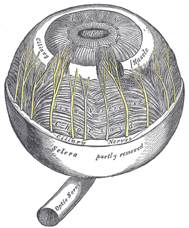
{Public domain illustration depicting ciliary muscle via Wikipedia.}