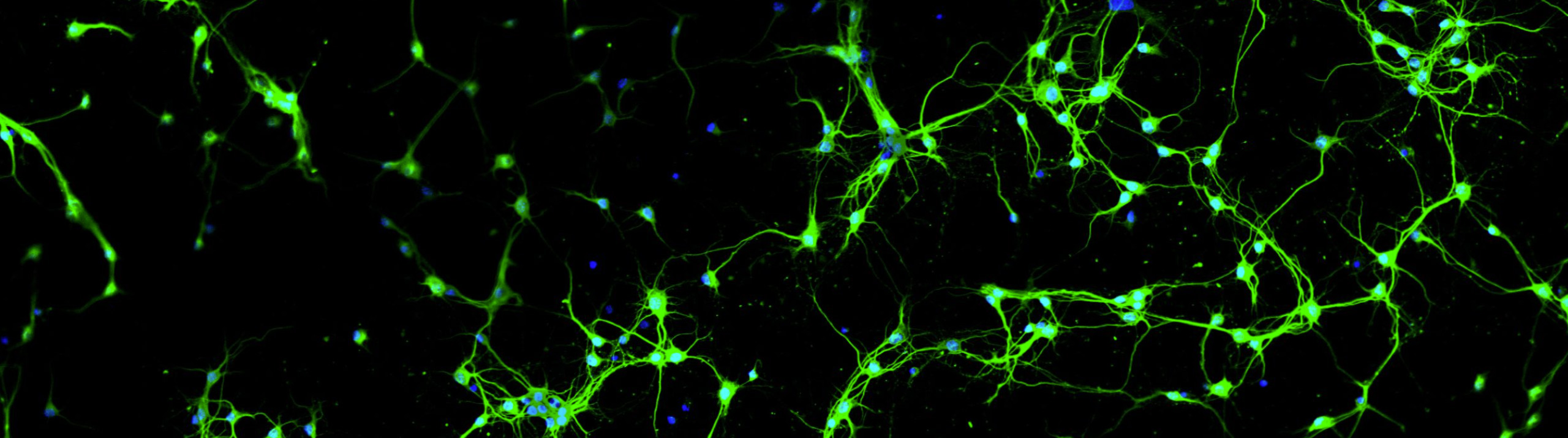
Columbia Scientists Map Neocortical Circuit Connectivity with Neurolucida
A willowy pair of pyramidal cells engage in an intricate dance with a dense mass of basket cells on the cover of the September 14, 2011 issue of the Journal of Neuroscience.
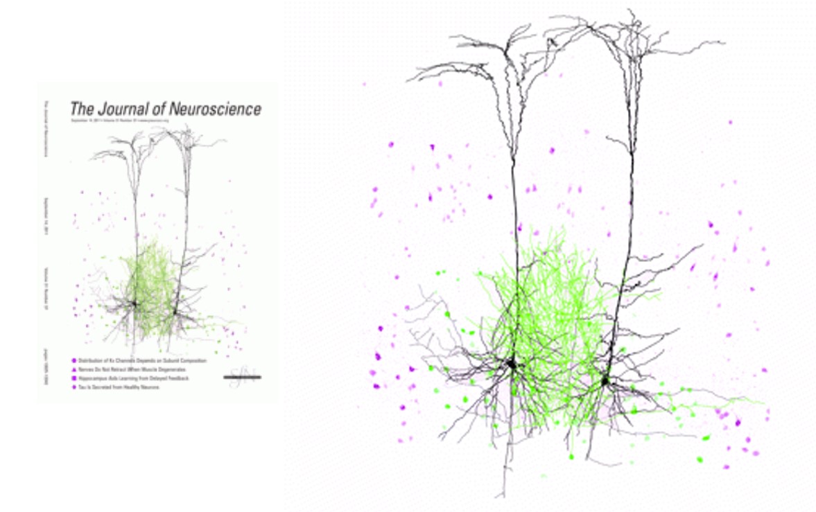
This exquisite image illustrates recent work by Columbia University researchers Dr. Adam M. Packer and Dr. Rafael Yuste, who used Neurolucida to study circuit connectivity in the mammalian neocortex.
According to the paper “Successfully filled and stained neurons were reconstructed using Neurolucida software (MicroBrightField). The neurons were viewed with a 100× oil objective on an Olympus IX71 inverted light microscope or an Olympus BX51 upright light microscope. The Neurolucida program projected the microscope image onto a computer drawing tablet. The neuron’s processes were traced manually while the program recorded the coordinates of the tracing to create a digital, three-dimensional reconstruction. The x- and y-axes formed the horizontal plane of the slice, while the z-axis was the depth. The user defined an initial reference point for each tracing. The z-coordinate was then determined by adjustment of the focus. In addition to the neuron, the pia and white matter were drawn. Axon and dendrite densities were calculated from the Neurolucida reconstruction using the TREES toolbox (Cuntz et al., 2010). The densities were calculated with voxels 5 μm on each side.” (Packer, Yuste, 2011)
Read the open access article in The Journal of Neuroscience.
Packer, Yuste. “Dense, Unspecific Connectivity of Neocortical Parvalbumin-Positive Interneurons: A Canonical Microcircuit for Inhibition?” The Journal of Neuroscience, 14 September 2011, 31(37):13260-13271; doi:10.1523/JNEUROSCI.3131-11.2011