Toronto Scientists Get First Direct Measurement of Myometrial SMCs in Pregnant Rat Uterus With Stereo Investigator
During pregnancy, the uterus grows to accommodate the increasing size of the fetus within. Makes sense. But what is it exactly that compels the uterus to get bigger? If you said pregnancy hormones, you’re right. And if you said the growing fetus, stretching the uterine walls, you’re right too.
Researchers attribute the growth of the uterus during pregnancy to both hormones and mechanical stretch (the fetus pressing on the uterine walls). In early pregnancy, the smooth muscle cells (SMCs) in the middle layer of the uterine wall (myometrium) increase in number, causing the uterus to grow larger. And as the pregnancy progresses, these cells actually get bigger. Scientists say the pressure of the fetus pushing on the uterus causes this increase in growth, but until now, there hasn’t been any accurate numerical data to uphold these claims.
Previous studies used the protien:DNA ratio as a marker for cellular hypertrophy, but it is speculated that this technique may not reflect the true degree of muscle cell growth during gestation.
Scientists at Mount Sinai Hospital’s Samuel Lunenfeld Research Institute in Toronto examined uterine tissue from non-pregnant rats, normally pregnant rats, and rats only pregnant in one of the two horns found within the uterus.
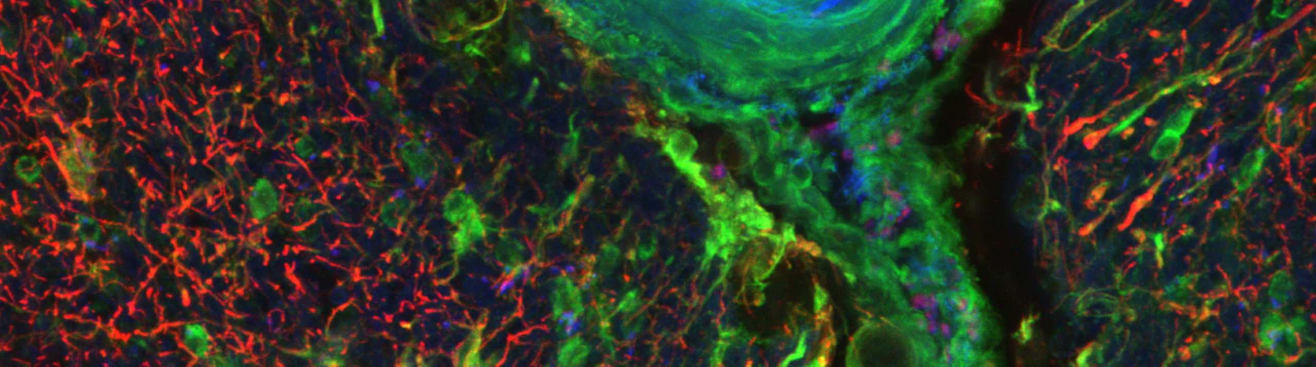
Led by Dr. Oksana Shynlova, the research team used Stereo Investigator and the point sample intercept probe to conduct a thorough stereological analysis of smooth muscle cell volume in uterine tissue. They gathered direct data on the diameters of individual cells marked with immunohistochemical staining, and collected an unbiased average cell volume for each stage of gestation.
Their study confirmed low cell growth in early pregnancy, with an increase in cell size as gestation continued, and a threefold increase in muscle cell volume toward the end of the term. Since the cellular hypertrophy was only observed in the horn containing a fetus, the researchers suggest that mechanical stretch may be the signal for these cells to grow larger, and not hormones alone.
“Although we and other investigators have shown earlier hypertrophic changes of uterine myocytes, this study is the first to directly measure the volume of myometrial SMCs throughout pregnancy,” the authors explained in their paper “Mechanical stretch regulates hypertrophic phenotype of the myometrium during pregnancy.”
Oksana Shynlova, Ruth Kwong, and Stephen Lye, “Mechanical stretch regulates hypertrophic phenotype of the myometrium during pregnancy” 2009 (Reproduction 2010;139:247.)
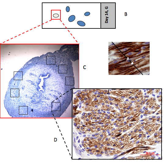
{Image: Using Masking tool the area of interest (myometrium or decidua in each slide)(B) containing 4-6 uterine biopsies, A) is chosen. Then the software program randomly selects 2% of the total area representing 25-45 images per each slide. (C) In each image the stereology software applies 4 counting frames. (Courtesy of the Samuel Lunenfeld Research Institute)}
For the latest news about MBF Bioscience and our customers, fan us on Facebook and follow us on Twitter